39+ Left Adnexal Mass Ultrasound Pics. Incidental adnexal masses diagnosed on ultrasound, ct or mri performed for an unrelated reason have increased in frequency with increased use of when an incidental adnexal mass is identified, further management will depend on whether the lesion is clearly benign or malignant, or indeterminate. Thanksnarrative pelvic ultrasound ** history **:
Malignant masses, us features of invasive ovarian cancer, ovarian.
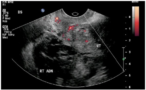
This is what is called the adnexa region that being said, your doctor may want to monitor the situation with regular ultrasounds and pelvic exams. One of the most important factors used to determine the clinical suspicion of malignancy of an adnexal mass is the sonographic appearance. Rlq ultrasound, onion skin structure? Key decision points are menopausal status, presence of concerning ultrasound findings, and.